
For further information, please watch the video: https://www.youtube.com/watch?v=EJBOxRt-dIg
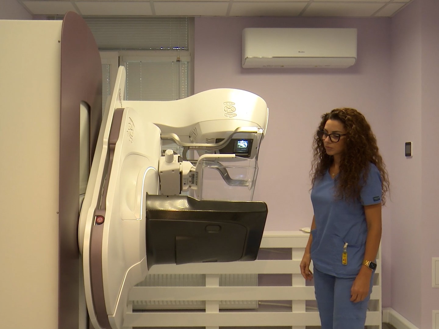
MU–Varna is investing in a latest-generation mammography system, which will be used for scientific research in the field of breast cancer diagnosis and screening. The equipment is expected to enable scientists to study and propose new techniques for early detection of breast cancer. The new acquisition is the result of the activities of scientific group “Early Diagnosis and Prevention of Oncological Breast Diseases by Using New Technologies” (ELPIDA) under the project “Enhancement of Translational Excellence Achievement in Medicine (MUVE-TEAM)” of MU–Varna, funded by the European Union.
The equipment will foster the development of experimental models and protocols that in the future could be applied to patients in the field of breast cancer diagnosis and screening. “The mammography unit will also be used in examinations with the so-called phantoms—these are test samples that allow specialists to take numerous measurements,” explained Assoc. Prof. Eng. Zhivko Bliznakov from the Department of Medical Equipment, Electronic and Information Technologies in Healthcare.
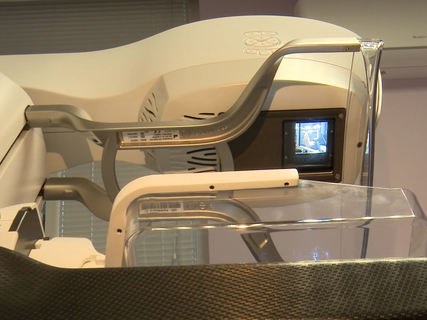
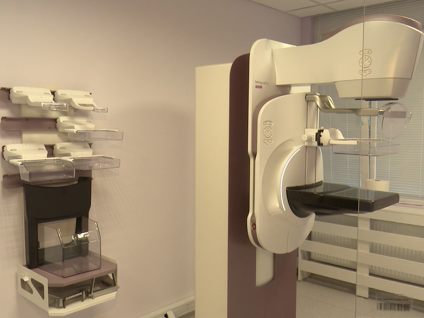
According to specialists, the new equipment is a latest-generation mammography system that makes possible the performance of specific examinations. Besides standard 2D examination, the apparatus also allows for detailed presentation of the findings in 3D examination – tomosynthesis, which provides extremely accurate diagnostics, especially in patients with high breast density.
The innovative medical technology also allows for contrast-enhanced mammography. This method combines standard mammography with an image of the contrast agent, which highlights areas with increased blood supply, characteristic of tumour formations. It is particularly useful in women with dense mammary glands, in cases of unclear findings from other examinations, or when magnetic resonance imaging (MRI) is not an option.
“For a socially significant disease such as breast cancer, mammography is crucial for monitoring and screening benign and malignant findings in the mammary glands,” underlined Assoc. Prof. Dr. Chavdar Bachvarov, Head of the Department of Diagnostic Imaging, Interventional Radiology and Radiotherapy at MU-Varna. The specialist commented that this would allow the University scientists to make a contribution to early diagnosis of breast cancer.